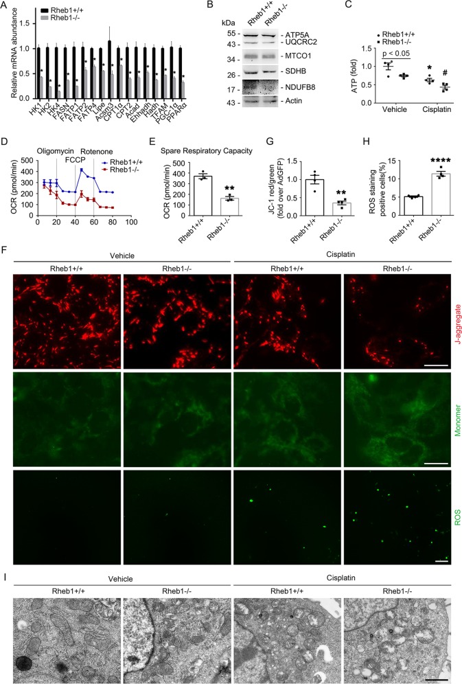
Fig. 6. Ablation of Rheb1 in primary cultured tubular cells leads to mitochondrial defect.
a Real-time qRT-PCR analysis showing the mRNA abundance of mitochondrial related genes for primary cultured tubular cells. b Western blot assay showing the abundance of mitochondrial respiratory chain complexes in primary cultured tubular cells. c The graph showing the ATP level in primary cultured tubular cells with or without cisplatin treatment. *P < 0.05 vs. Vehicle control, #P < 0.05 vs. Ad-GFP-infected cells treated with cisplatin, n = 4. d, e The graph showing the results of OCR assay, **P < 0.01, n = 4. f Representative images of DCFH-DA staining for ROS (Scale bar = 20 µm) and JC-1 staining (Scale bar = 5 µm). g, h Quantitative analyses of JC-1 fluorescence intensity (red/green) and DCFH-DA -staining positive cells after cisplatin treatment. Data is presented as the percentage of the staining positive cells. *P < 0.05 vs. vehicle control, #P < 0.05 vs. Ad-GFP-infected cells treated with cisplatin, n = 4. i Representative transmission electron micrographs of the primary cultured tubular cells infected with Ad-Cre and Ad-GFP with or without cisplatin treatment as indicated. *P < 0.05 vs. vehicle control. (Scale bar = 2 µm). HK1, 2, and 4: hexokinase 1, 2, and 4; FASN: fatty acid synthase; FATP1, 2, and 4: fatty acid transport protein 1, 2, and 4; HSL or Lipe: hormone-sensitive lipase; Acsm3: acyl-CoA synthetase medium-chain family member 3; Cpt1α and 2: carnitine palmitoyl transferase 1α and 2; Acad acyl-Coenzyme A dehydrogenase; Ehhadh enoyl-coenzyme A hydratase/3-hydroxyacyl-coenzyme A dehydrogenase; Hadh hydroxyacyl-Coenzyme A dehydrogenase; TFAM mitochondrial transcription factor A; PGC-1α: peroxisome proliferator-activated receptor γ coactivator-1α; PPARα: peroxisome proliferators-activated receptor α.

