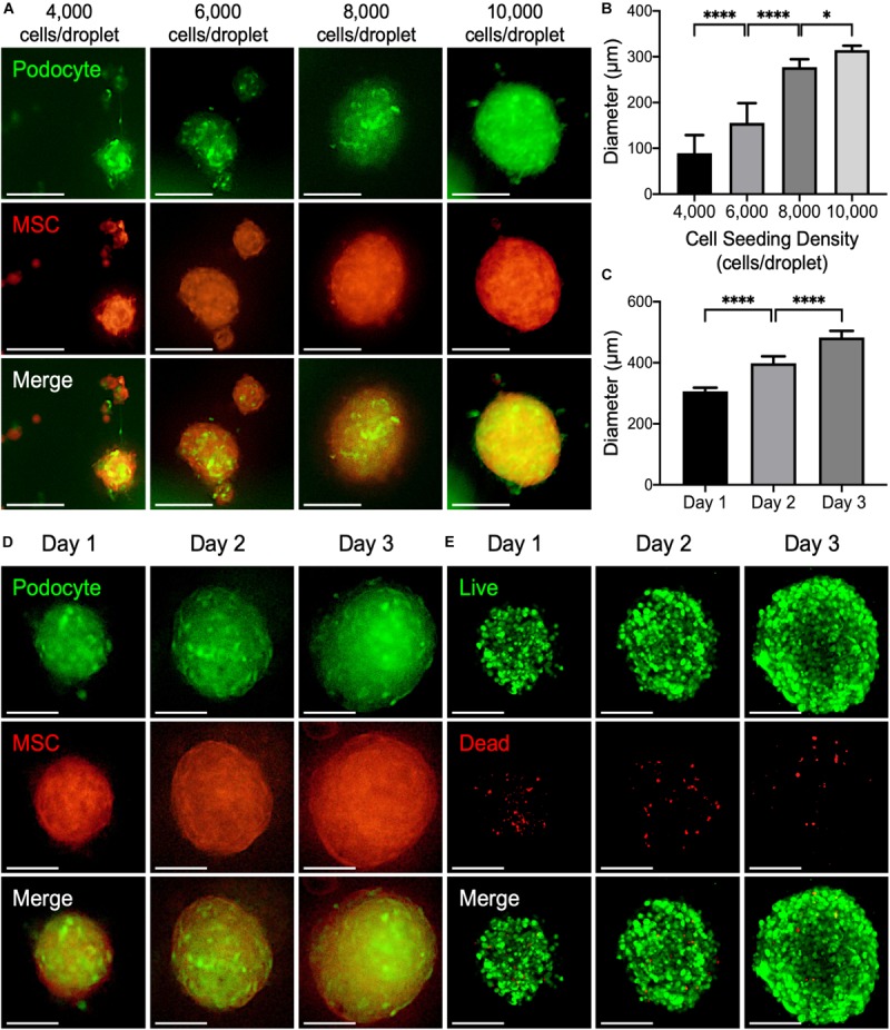
FIGURE 4.

(A) Representative fluorescence images of 3D kidney microtissues comprised of podocyte (green) and mesenchymal stem cells (MSCs; red) fabricated using a PANDA chip. The microtissues were formed within 24 h. (B) The diameters of the formed 3D microtissues after 1 day of culture (*P < 0.05; ****P < 0.001; N = 20). (C) The diameters of the formed 3D microtissues cultured on different days (****P < 0.001. N = 20). (D) Representative fluorescence images of 3D kidney microtissues cultivated for various periods. (E) Confocal images of live (green) and dead (red) cells. All scale bars represent 200 μm.
