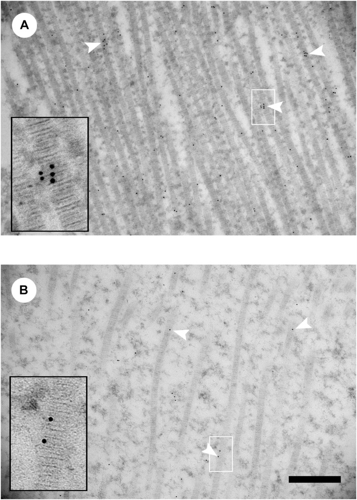
FIGURE 3.
Decorin immunogold transmission electron microscopy images of collagen fibrils from wooden breast-affected muscle of Line A (A) and Line B (B). White arrowheads indicate gold particles labeling collagen fibrils. Insets show enlarged images of gold labeling individual collagen fibrils in the region indicated by the white box. Scale bar = 500 nm. (Figure reproduced from Tonniges et al. (2019). Avian Dis. 63, 48–60).

