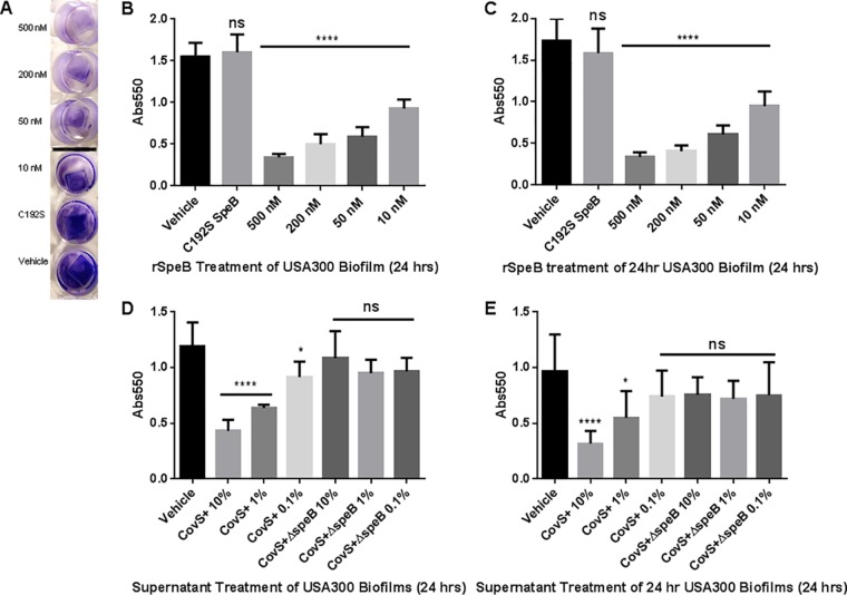
FIG 2.
Biofilm density after SpeB treatment measured by crystal violet assay. (A) Representative image of dose-dependent reduction in biofilm density after treatment with r-SpeB. (B) Crystal violet assay of biofilm formation over 24 h with r-SpeB. (C) Crystal violet assay of the degradation of a 24-h-old biofilm after an additional 24-h treatment with r-SpeB. (D) Biofilm formation over 24 h in the presence of supernatants of AP53CovS+ and AP53CovS+ ΔspeB GAS strains. (E) Biofilm degradation of 24-h-old biofilm over an additional 24 h with GAS supernatants. *, P < 0.05; ****, P < 0.0001; ns, not significant.

