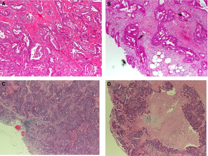
FIGURE 6.

Hematoxylin–eosin staining of tumor samples. Shown is the staining of (A) the primary tumor, (B) metastatic peritoneal tumor, (C) patient‐derived xenograft (PDX) from the primary tumor, and (D) PDX from the metastatic peritoneal tumor. The pathological evaluation shows the presence of moderately differentiated tubular adenocarcinoma in the PDX, which is similar to that in the original tumor. Magnification (40×)
