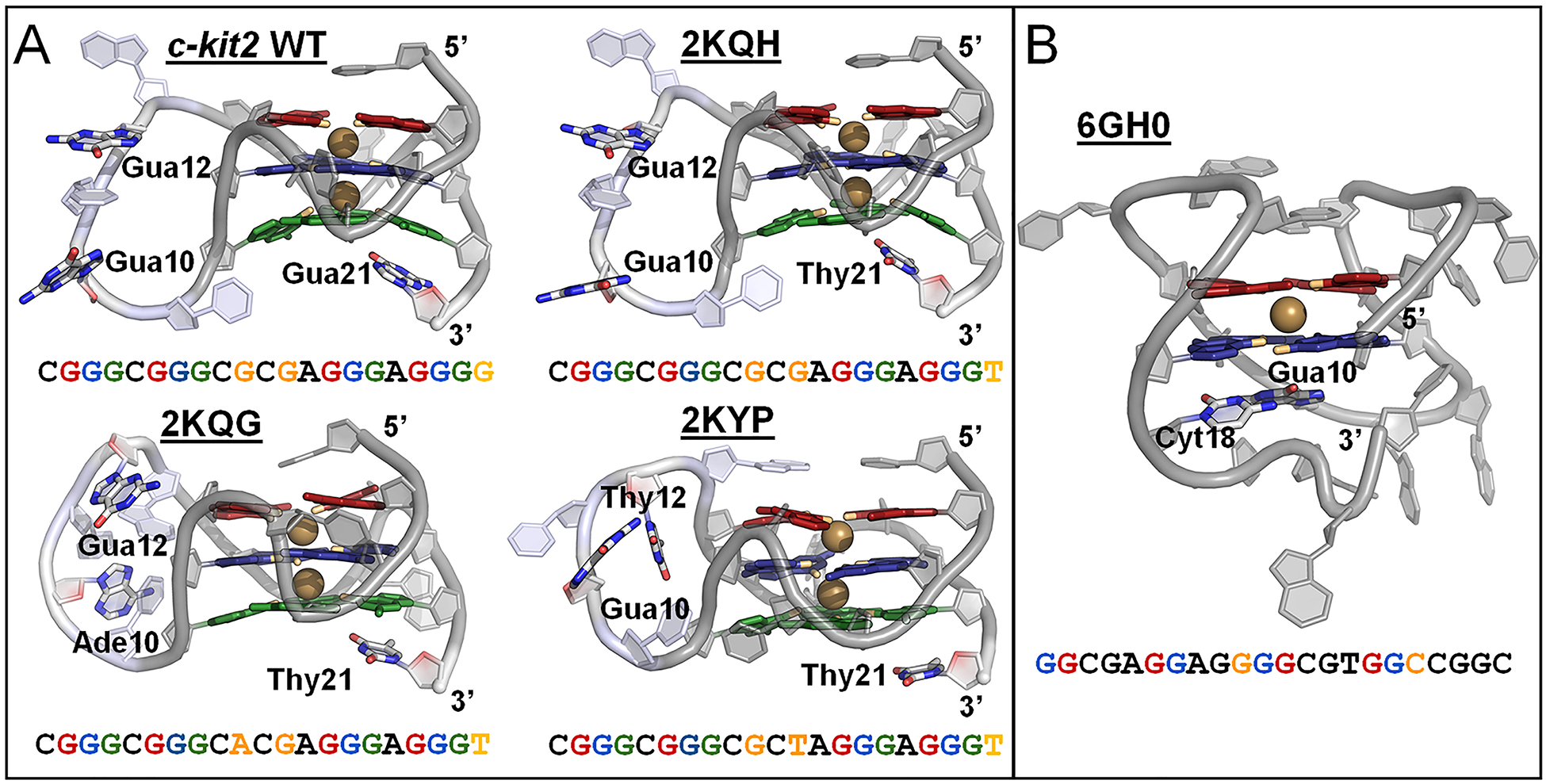
Figure 2.

Structure and sequence of the c-kit2 and c-kit* GQs. (A) Cartoon representation of starting c-kit2 structures, highlighting bound K+ ions (colored gold) and nucleotides of the long propeller loop (residues 9–13; colored light blue). Residues 10, 12, and 21 vary in the c-kit2 structures and are represented as colored sticks and orange text. (B) Cartoon representation of starting c-kit* structure, highlighting a bound K+ ion (colored gold) and structurally important linker nucleotides (Gua10 and Cyt18) in colored stick representations and orange text. The guanine bases of the GQ cores are colored by tetrad: 1 – red, 2 – blue, and 3 – green (in c-kit2 GQs). O6 atoms pointing inward to coordinate K+ are colored orange.
