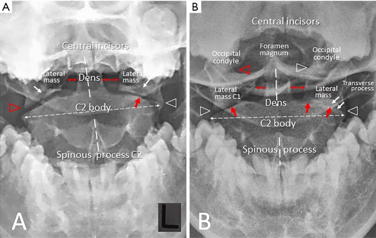
Figure 2.
Odontoid view comparison of pre-and-post intervention. (A) Initial radiograph demonstrated mal-alignment of the right zygapophyseal (C1–C2) joint (red triangle), subchondral erosions in the lateral masses (white arrows), narrowing of the left C1–C2 joint (white arrow). In addition, tilt of the C2 median line, asymmetry of the C1 lateral masses and of the atlantodens intervals (double headed arrows) imply C1–C2 subluxation; (B) repeated radiograph showed asymmetrical size of the C1 lateral masses, displacement of the right lateral mass with respect to the alignment of occipital condyle (red triangle) and to the C2 facet, deteriorated changes of bilateral C1–C2 joints (red arrows) with subchondral sclerosis (white arrows), and reposition of the C2 median line.

