
Figure 1.
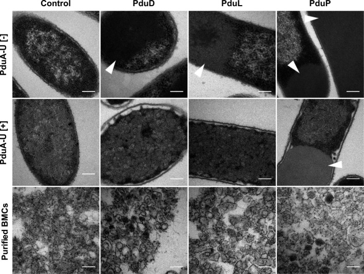
Encapsulation of native proteins in recombinant Pdu BMCs. Electron micrographs of Escherichia coli cells producing PduD, PduL, or PduP in the absence (top panel) and presence (middle panel) of a minimal BMC shell system (PduA‐U). The BMCs extracted from strains in the middle panel are also shown. The control sample contains an empty vector. Arrows indicate areas of protein aggregation. All scale bars are 200 nm