
Figure 1.
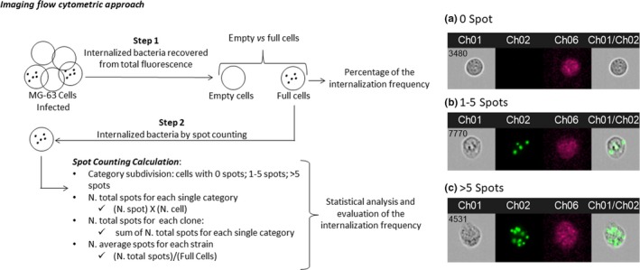
Images obtained by FlowSight® Imaging Flow Cytometer (Amnis® FlowSight® Millipore, Merck KgaA, Darmstadt, Germany), acquiring 10,000 events per sample: (a) MG‐63, without spots inside the cell; (b) a single MG‐63 cell with four spots inside (each spot was considered as a single bacterial cell); and (c) a single MG‐63 cell with >5 spots. CH1 brightfield; CH2 band nM 505–560; CH6 Side Scatter (SSC) 785 nM; CH1/CH2 merge brightfield and 505–560 nM band