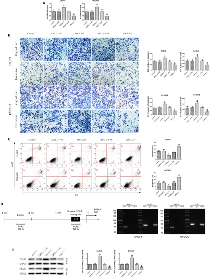
Figure 5.

CDC5L exerted an oncogenic role and regulated PEAK1 expression in CAOV3 and OVCAR3. A, CCK‐8 assay was used to determine the effect of CDC5L on proliferation of CAOV3 and OVCAR3 (data are presented as mean ± SD [n = 3, each group]; *P < .05 vs CDC5L (+)‐NC group; # P < .05 vs CDC5L (−)‐NC group). B, Effect of CDC5L on migration and invasion of CAOV3 and OVCAR3 (data are presented as mean ± SD [n = 3, each group]; *P < .05 vs CDC5L (+)‐NC group; # P < .05 vs CDC5L (−)‐NC group). Photographs were taken at 200× magnification. Scale bar represents 100 μm. C, Flow cytometry analysis of CAOV3 and OVCAR3 with altered expression of CDC5L (data are presented as mean ± SD [n = 3, each group]; *P < .05 vs CDC5L (+)‐NC group; ## P < .01 vs CDC5L (−)‐NC group). D, CDC5L bound to the promoter of PEAK1 in CAOV3 and OVCAR3. Transcription start site (TSS) was designated as +1. The putative CDC5L binding sites are indicated. Immunoprecipitated DNA was amplified by PCR. Normal rabbit IgG was used as negative control. E, Western blot analysis for CDC5L regulated expression of PEAK1 protein with GAPDH as endogenous control (data are presented as mean ± SD [n = 3, each group]; **P < .01 vs CDC5L (+)‐NC group; # P < .05 vs CDC5L (−)‐NC group)
