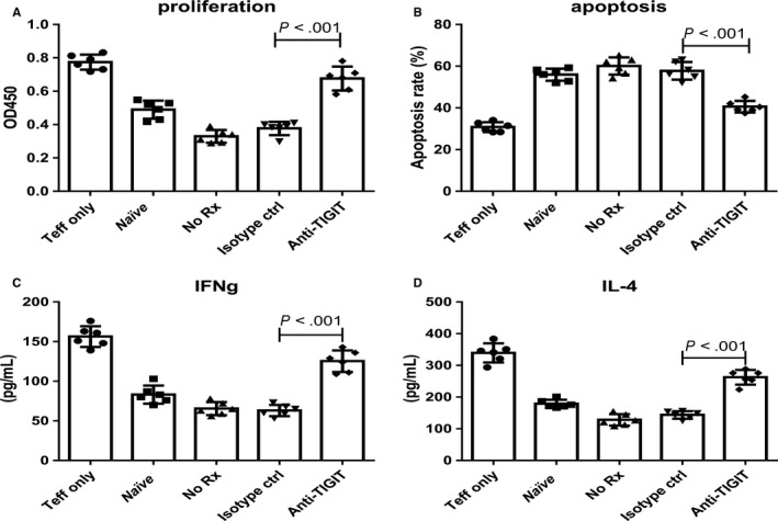
Figure 4.

Anti‐TIGIT treatment reduced immunosuppressive functions of CD4+ Tregs. Mice (six individuals per group) were injected i.p. with 1 × 10⁶ ID8 cells ten days before treatments. OC mice were injected thrice in 4‐d intervals with 100 μg of control or anti‐TIGIT mAb. Seven days after this, splenic CD4+ Tregs were isolated from different groups and were co‐cultured with normal CD4+ CD25− T effector cells from normal mice in the stimulation of anti‐CD3 (5 μg/mL) and anti‐CD28 (2 μg/mL) in a ratio of 1:1 for 24 h. Proliferation (A), apoptotic rate (B), and secretive ability (IFN‐γ and IL‐4) (C and D) of CD4+ CD25− T effector cells were determined. “Naïve” indicates normal splenic CD4+ Tregs co‐cultured with normal CD4+ CD25‐ T effector cells. Shown are the means ± standard deviation
