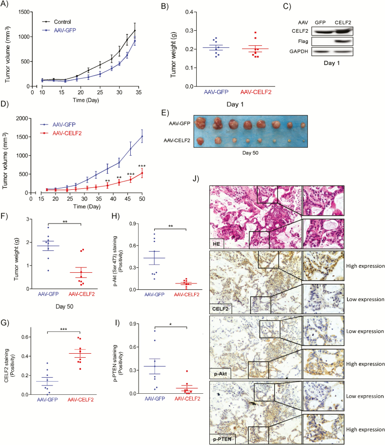
Figure 5.
AAV-mediated CELF2 protein expression inhibits tumor growth in a PDX mouse model. (A) Tumor growth kinetics over 36 days of uninfected or AAV-GFP-infected PDX. (B) Tumor weight at Day 1. (C) Western blot analysis of CELF2 protein expression in AAV-GFP- and AAV-CELF2-infected PDX tissues at Day 1. (D) Tumor growth kinetics over 50 days of AAV-GFP- (as control) or AAV-CELF2-infected PDXs. Tumor size was measured twice a week and calculated based on the formula: length × width2 × 0.5. (E) Tumor size and (F) tumor weight at Day 50. (G) CELF2, (H) pAkt (Ser473) and (I) p-PTEN expression and HE staining in harvested xenograft tissues were assessed by immunohistochemistry. The integrated optical density was evaluated using the Image-Pro premier software offline (v9.0) program. (J) Representative photographs of immunohistochemistry analysis for each antibody are shown.

