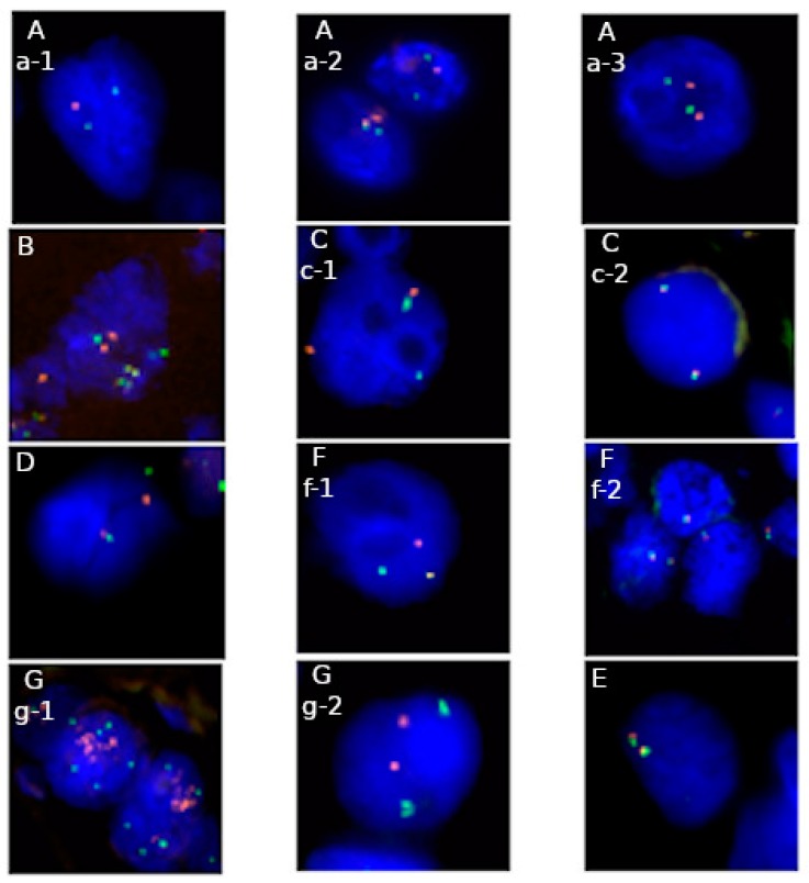
Figure 1.
Applications of fluorescence in situ hybridization (FISH) in genetic diagnostics in solid tumors on FFPE material: A—1p/19q probe: a-1 deletion of 1p32 locus, a-2 normal signal pattern (cell on the left) and deletion of 19q13 locus (cell on the right), a-3 normal signal pattern (Abbott Molecular), B—dual fusion probe: fusions and normal signals pattern of COL1A1 and PDGFB loci (ZytoVision), C—break apart probe: c-1 rearrangement of ALK gene, c-2 normal signal pattern (Abbott Molecular), D—break apart probe: rearrangement of EWSR1 locus (Abbott Molecular), F—break apart probe: f-1 rearrangement of ROS1 gene, f-2 normal signal pattern (Empire Genomics), G—locus specific probe: g-1 amplification of HER2 locus, g-2 normal signal pattern (Abbott Molecular), E—break apart probe: normal signal pattern of SS18 locus (Abbott Molecular). Majority probes indicate region of interest in red color and control region—in green, excluding picture B, where red color indicates COL1A1 gene locus, green color—PDGFB gene locus, yellow color—fusion of COL1A1-PDGFB and PDGFB-COL1A1. Full names of available commercial probes are presented in Table 2.

