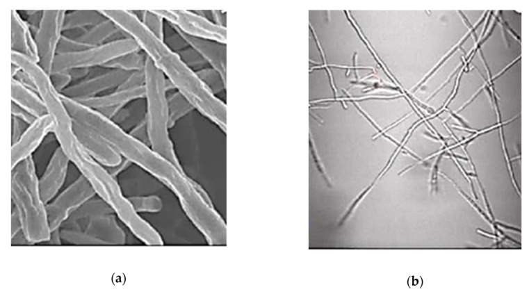
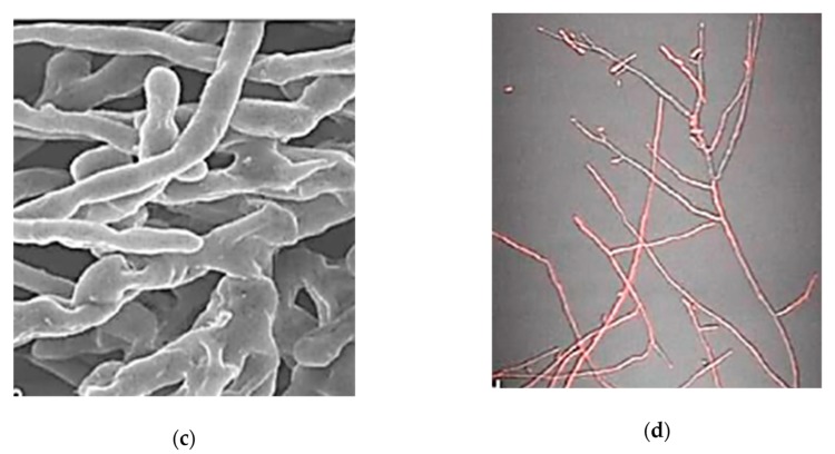
Figure 20.
Comparison of ultrastructural and cell permeability changes in F. oxysporum mycelium upon chitosan bionanomaterial (BNM) exposure: (a) and (c) show ultrastructural changes determined by SEM analysis of F. oxysporum mycelium of control (a) and chitosan BNMs (400 µg/mL) (c); (b) and (d) show cell permeability analysis data by propidium iodide assay; (b) influx of control; (d) influx of chitosan BNMs treatment (modified after [84]).


