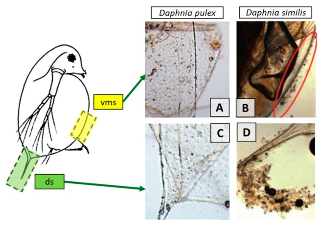
Figure 29.
Representative image of distal spine (ds) and ventral margin of the shield (vms) in Daphnia pulex and D. simillis exposed to 10 mg/L of CeO2 NPs for 48 h. Note the accumulation of particles onto the cuticle of D. simillis. (Modified after [146]).

