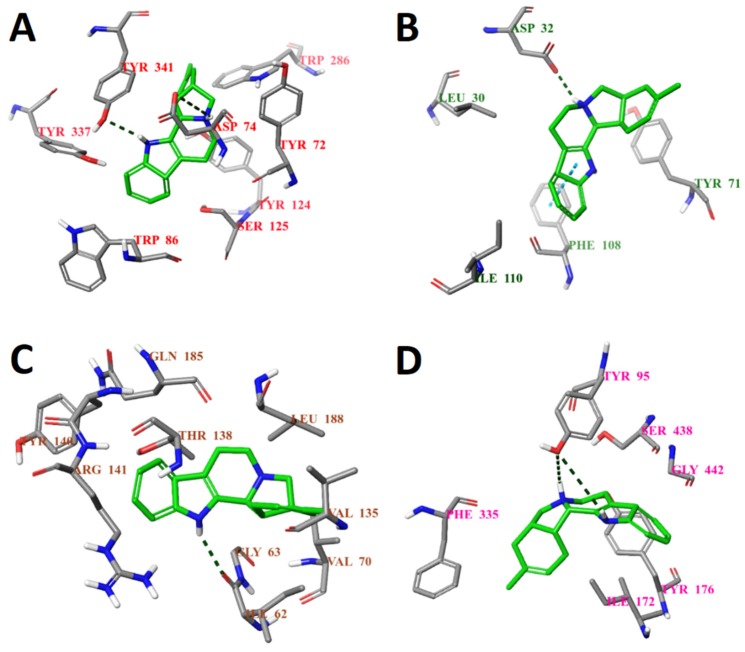
Figure 5.
Calculated binding modes of compound ZINC4027357 (A) in the active site of AChE (ID: 4EY6); (B) in the active site of BACE1 (ID: 6EQM); (C) in the active site of GSK3β (ID: 1PYX); (D) in the central active site of SERT (ID: 5I6X). The amino acid residues of target proteins are colored as gray (carbon), blue (nitrogen), red (oxygen), and white (hydrogen). Hydrogen bonds formed between compound and residues of target proteins are represented by green dashed lines, and pi–pi stacking is represented by a blue dashed line.

