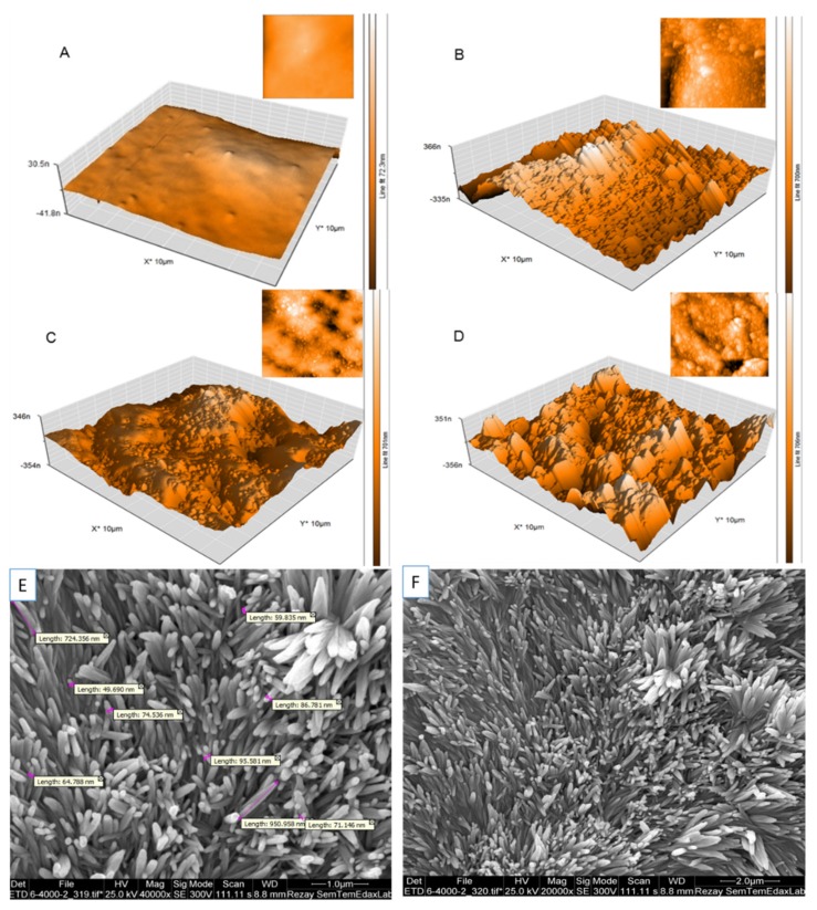
Figure 2.
Substratum atomic force microscopy (AFM) topography of unmodified (A) and modified PDMS surfaces with nanoparticle coating: GO (B), GQD (C), SPION (D), recorded in tapping mode. The contrast covers height variations in the 0–30 nm scale in A and in the 0–350 nm scale in B, C, and D. Scanning electron microscopy image of the morphology of gold nanowires mixed up with PDMS (E,F).

