Fig. 7.

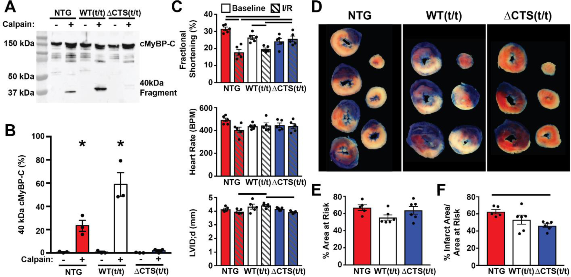
Prevention of calpain proteolysis of cMyBP-C reduces infarct size following ischemia-reperfusion injury. (A) Western blot of cMyBP-C from NTG, WT(t/t), and ΔCTS(t/t) myofilaments incubated with and without calpain in 1.25 mM calcium shows the generation of the N’-terminal 40 kDa cMyBP-C proteolysis fragment. The fragment appears at a higher weight in the WT(t/t) sample due to the presence of a Myc tag. (B) Quantification of the percent of 40kDa cMyBP-C fragment to total cMyBP-C (n = 3) (* P<0.05; two-way ANOVA). (C) Echocardiography results from NTG, WT(t/t), and ΔCTS(t/t) mice pre- and post-I/R show preserved systolic function in ΔCTS(t/t) mice but not in NTG or WT(t/t) controls. Additionally, NTG and WT(t/t) hearts showed ventricular wall thinning following I/R, whereas this was not apparent in ΔCTS(t/t) hearts (n=5) (horizontal black bars represent p<0.05 between indicated groups; two-way ANOVA). (D) Example tissue sections of NTG, WT(t/t), and ΔCTS(t/t) hearts stained with TTC and Phthalo Blue pigment solution following I/R injury. After cutting into cross sections, tissue mass, area at risk, and infarct areas were measured. Blue myocardium represents the remote area, red stained myocardium represents the area-at-risk, and the white regions are infarcted tissue. (E) Quantification of these areas show the area at risk was similar between the three groups. (F) The % infarct area was significantly reduced in ΔCTS(t/t) hearts. (n = 5, NTG; 6, WT(t/t); 6, ΔCTS(t/t)). Horizontal black bar represents p<0.05 between indicated groups; one-way ANOVA). All data are mean ± SEM.
