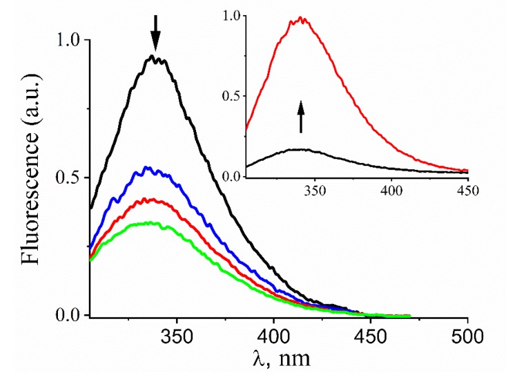
Figure 1.
Changes in the intrinsic fluorescence of Prx1C83SC173S upon S-nitrosation and denitrosation. Representative emission spectra (λexc. = 280 nm) of reduced Prx1C83SC173S (5 µM) before (black trace) and 2 (blue trace), 5 (red trace), and 10 min (green trace) after addition of GSNO (nitrosoglutathione) (400 µM). Inset: representative emission spectra (λexc. = 280 nm) of S-nitrosated Prx1C83SC173S (5 µM) before (black trace) and 10 min (red trace) after addition of glutathione (400 µM). The rows indicate the temporal spectral changes. The incubations were performed in phosphate buffer (50 mM) containing DTPA (diethylenetriaminepentaacetic acid) (0.1 mM), pH 7.4 and 25 °C.

