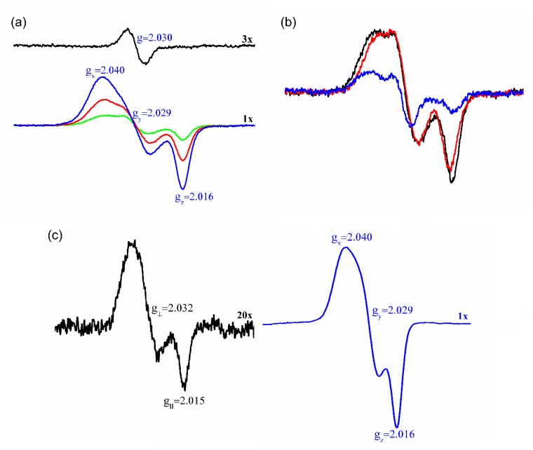
Figure 5.
EPR detection of dinitrosyl-iron complex (DNIC)-Prx1 complexes. (a) Representative EPR spectra of DNIC-GS (60 μM) before (black trace) and 30 min after addition of 30 (green trace), 60 (red trace) or 120 μM (blue trace) Prx1 scanned at room temperature. (b) Representative EPR spectra of DNIC-GS (60 μM) 30 min after addition of 30 μM of wild-type Prx1 (black trace), Prx1C83SC173S (red trace) or Prx1C52S (blue trace) scanned at room temperature. (c) Time-dependent 77K EPR spectrum of DNIC-GS (60 μM) plus Prx1 (60 μM) after 2 (black trace) or 30 min (blue trace) incubation. Aliquots of the incubation were removed at the specified times and frozen to stop the interaction and the EPR spectrum acquired at 77 K. All the incubations were performed in phosphate buffer (50 mM) containing DTPA (0.1 mM), pH 7.4 (final) at 25 °C.

