Figure 7.
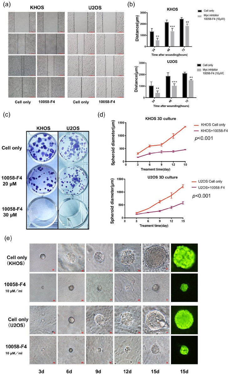
Inhibition of Myc suppressed osteosarcoma cell migration, clonogenicity, and spheroid growth. (A) Relative migration distance of KHOS and U2OS cells at different time points (0, 24, 48, and 72 h) when treated with the Myc inhibitor 10058-F4. (Original magnification value, ×100. Scale bar 1000 µm). (B) Quantification of cell migration distance of KHOS and U2OS cells after 10058-F4 treatment. **p < 0.01, ***p < 0.001 compared with the untreated control group. (C) Representative images of KHOS and U2OS cell colony formation after treatment with 10058-F4 at different concentrations (0, 20, 30 µM) for 15 days. (D) Spheroid diameters of KHOS and U2OS cells cultured in 3D gels. p < 0.001 compared with the untreated control group. (E) Representative images of osteosarcoma spheroids after treatment with the Myc inhibitor at different time points (3, 6, 9, 12. and 15 days). Original magnification, ×200. Scale bar 100 µm.
3D, three-dimensional; Myc, avian myelocytomatosis viral oncogene homolog.

