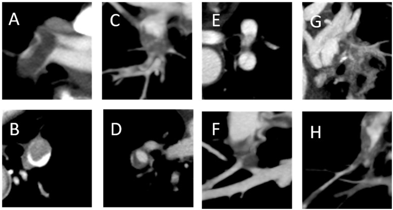
Fig. 1.
Acute thrombus (a and b) and chronic thrombus (c–h) according to CT. (a) Well-defined central filling defect; (b) eccentric filling defect with acute angles within the vessel wall; (c) pouch defect; (d) filling defect with obtuse angle within the vessel wall (crescent shape); (e) band lesion; (f and g) web lesion with calcification; and (h) abrupt vessel narrowing with irregular intima.

