Figure 1.
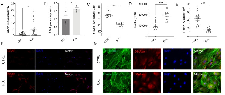
Reactive astrocytosis results in cytoskeletal remodeling. (A) anti-glial fibrillary acid protein (GFAP) immunoreactivity was quantified by fluorimetry. A statistically significant increase in GFAP immunoreactivity was observed in activated astrocytes compared to control (n = 20–21; p < 0.01). (B) Quantitative immunoblot analysis revealed a statistically significant increase in GFAP protein expression in activated astrocytes (n = 3; p < 0.05). (C) Quantification of individual F-actin fiber lengths revealed a statistically significant 36% ± 2% decrease in activated optic nerve head astrocytes (n = 10; p < 0.001). (D) A statistically significant 87% ± 12% increase in G-actin immunofluorescence was observed in activated astrocytes (n = 10; p < 0.001). (E) The F-actin to G-actin ratio showed a statistically significant decrease in activated astrocytes (n = 10, p < 0.001). (F) Representative images of GFAP immunocytochemistry. (G) Representative images of actin staining. Oligomeric F-actin and monomeric G-actin were visualized using ActinGreenTM ReadyProbesTM Reagent and Alexa FluorTM 594 conjugated DNAseI, respectively. Data were analyzed by Student’s t-test; * p < 0.05, ** p < 0.01, *** p < 0.001. Scale bar: (F) 25 μm, (G) 10 µm. CTRL = control; R.A. = reactive astrocytosis.

