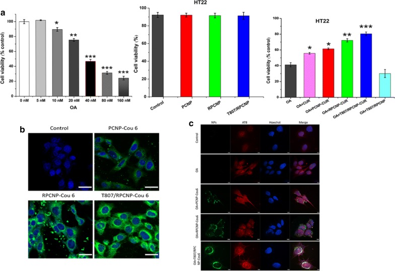
Fig. 3.
Cytotoxicity effect and specific tau targeting capability of biomimetic formulations. a The suitable concentration of OA for the model of AD after exposure to different concentration for 12 h, then the toxicity of different NPs on HT22 cells treated with or without 40 nM OA at a higher concentration. Error bars represent standard deviation. Statistical analysis was performed using a one-way ANOVA test, with *** indicating p < 0.001, ** indicating p < 0.01, and * indicating p < 0.05 (n = 6 per group). b The internalization of COU-tagged biomimetic NPs in HT22 cells (scale bar = 20 μm). c Confocal fluorescence images of different NPs in OA-treated HT22 cells after 4 h incubation. Cells stained with hoechst33258 are shown in blue fluorescence. The immunofluorescence staining of OA-treated HT22 cells with phosphor-tau AT8 (ser202, Thr205) antibody are shown in red fluorescence. COU-labeled PCNP, RPCNP and T807/RPCNP are shown in green fluorescence (scale bar = 50 μm)

