Figure 6.

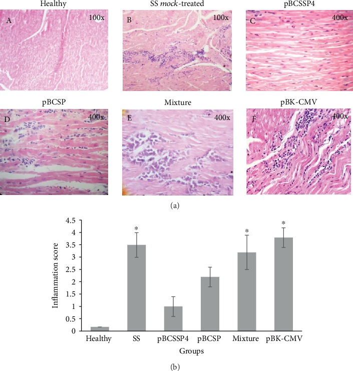
Histological ventricular myocardium findings of T. cruzi-infected dogs treated with DNA vaccines. (a) Representative micrographs of the heart tissue from all groups are shown. (A) Transverse section of the LV myocardium showing healthy tissue. (B) LV myocardial cross section of the SS mock-treated infected control group showing severe multifocal coalescent lymphoplasmacytic myocarditis (score: 4). (C) Longitudinal section of the LV myocardium of the pBCSSP4 plasmid-treated group showing mild multifocal interstitial lymphoplasmacytic myocarditis (score: 2). (D) Longitudinal section of the LV myocardium of the pBCSP plasmid-treated group showing mild multifocal interstitial lymphoplasmacytic myocarditis (score: 2) and moderate degeneration of muscle fibers. (E) RV myocardial longitudinal section of the plasmid mixture-treated group showing lymphocytic moderate multifocal coalescent myocarditis (score: 3). (F) Longitudinal section of the LV myocardium of the empty vector plasmid-treated group showing moderate to severe multifocal coalescent myocarditis (score: 4). Hematoxylin and eosin staining. (b) Inflammatory lesion (inflammatory cell infiltrates) scores. Data are expressed as the mean ± S.D., and differences were considered significant when ∗P ≤ 0.05 by the Kruskal-Wallis test for the healthy group versus the SS mock-treated, pBCSSP4, pBCSP, mixture, and pBK-CMV groups.
