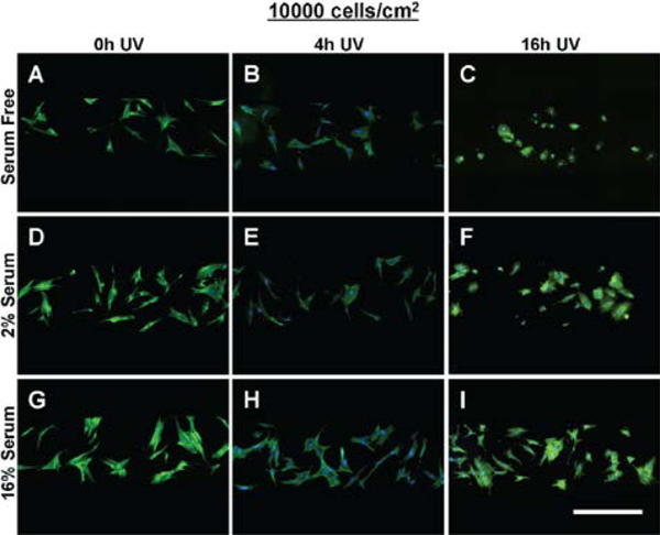
Figure 5:
The morphology of MSCs seeded at 5000 cells/cm2 within tctPS microchannels grown in (A-C) serum free, (D-F) 2% serum, and (G-I) 16% serum conditions is shown for tctPS substrates that were previously subjected to (A, D, G) 0h, (B, E, H) 4h, and (C, ,G, I) 16h of UV exposure prior to cell seeding. MSCs were stained for actin (green) and nuclei (blue). Scale bar is 500 microns.

