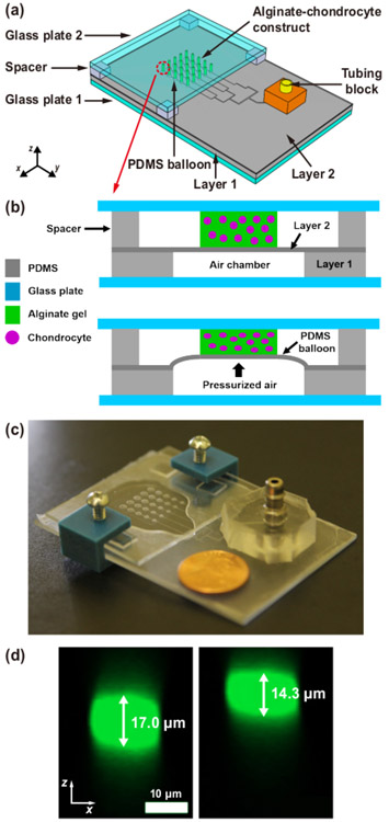
Figure 1. Microfluidic chondrocyte compression device.
(a) Schematic of the assembled device. A 5 × 5 array of alginate–chondrocyte constructs are aligned on PDMS balloons with 5 different diameters (D = 1.2, 1.4, 1.6, 1.8 and 2.0 mm), where D is the diameter of PDMS balloon (or air chamber). (b) Schematic of the device operation. The device is actuated by pneumatic pressure which expands PDMS balloons. (c) Image of an actual device (coin diameter = 19 mm). (d) Vertical cross-sections of a chondrocyte before (left) and under (right) compression on the largest PDMS balloon (D = 2.0 mm) (cell compressive strain, εcell = ∣cell height change/initial cell height∣ × 100 = 16%). This figure is reproduced from 26.

