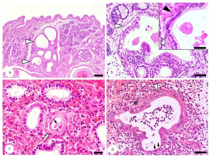
Figure 4.
Equine endometrium, endometrosis, microscopic features, hematoxylin-eosin stain: (a) At low magnification, endometrotic glands (arrowheads) are recognized by periglandular fibrosis that is often associated with their cystic dilation, irregular shape, and/or nesting. Bar = 100 µm. (b) Nested endometrotic glands surrounded by inactive (white arrowheads) and active fibrosis (black arrowhead, inset). Inactive stromal cells have elongated hyperchromatic nuclei (white arrowheads), whereas activated stromal cells show oval hypochromatic nuclei (black arrowheads). Bar = 40 µm. Bar inset = 20 µm. (c) Mild inactive destructive endometrosis of a single gland (white arrow): In the destructive form, lining epithelia show degeneration/necrosis and intraluminal desquamation (thin arrow). Bar = 20 µm. (d) Concurrent presence of destructive endometrosis and endometritis: Periglandular fibrosis is labelled by a white arrowhead, and degeneration and attenuation of lining epithelia are labelled by black arrows, and inflammatory cells within the stroma are labelled by an asterisk. Bar = 50 µm.

