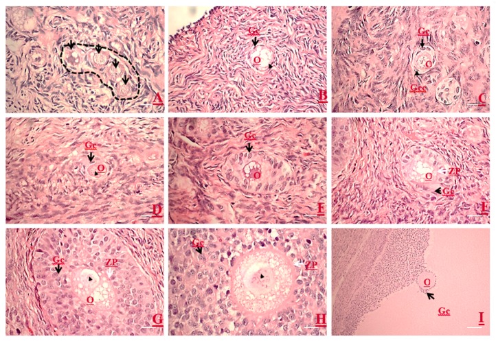
Figure 1.
Microscopic morphologies of growing oocytes, follicles, and the associated follicular cells at different stages of folliculogenesis by using semi-thin sections of porcine ovaries. (A) A cluster of primordial follicles (enclosed in dot line) is observed in the cortex area. (B) Note the flattened granulosa cells (Gc) (arrow) are surrounding an immature oocyte, and the oocyte contains an eccentrically localized nucleus (arrowhead). (C) An activated primordial follicle: an oocyte is surrounded by two types of granulosa cells, i.e., cuboidal granulosa cells (arrow) at one pole and flattened granulosa cells (arrowhead). (D) A primary follicle oocyte is surrounded by a single layer of cuboidal granulosa cells (arrow), and the oocyte contains an eccentrically localized nucleus (arrowhead). (E–H) An early secondary follicle is transforming into a late stage secondary follicle with the onset of zona pellucida (ZP) formation (white arrow). Note that the increasing layers of cuboidal granulosa cells can be observed with no ZP structure (E), and all oocytes possess an eccentric germinal vesicle (GV nucleus, arrowhead). A very thin ZP (F) starts to form (white arrow). (G) The ZP is getting thicker as the granulosa layers increased. The granulosa cells start getting loosening while the ZP is thickening. (I) A tertiary follicle shows multiple layers of polar granulosa cells, antrum, and an eccentric cumulus–oocyte complex (COC). O, oocyte. Magnification: 100× (Scale bars, 100 µm) (A,B,D; E–G); 200× (scale bars, 50 µm) (C,H); and 40× (scale bar, 250 µm) (I).

