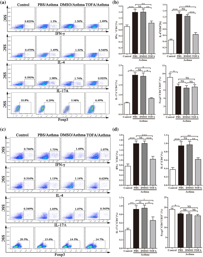
Fig. 5.
Reduced percentages of Th1, Th2, Th17 cells in TOFA-treated asthma mice. IFN-γ+, IL-4+, IL-17+ Th cells from CD4+ cells, Foxp3+ Th cells from CD4+CD25+ Th cells in lung tissue (a) and in spleen (c) were examined by flow cytometric analysis. The percentage of CD4+ cells expressing IFN-γ+, IL-4+, IL-17+ and the percentage of CD4+CD25+ cells expressing Foxp3+ in lung tissue (b) and in spleen (d). All data are represented as mean ± SEM (n = 5–6 mice per group). *p < 0.05; **p < 0.01; ***p < 0.001; NS, p > 0.05

