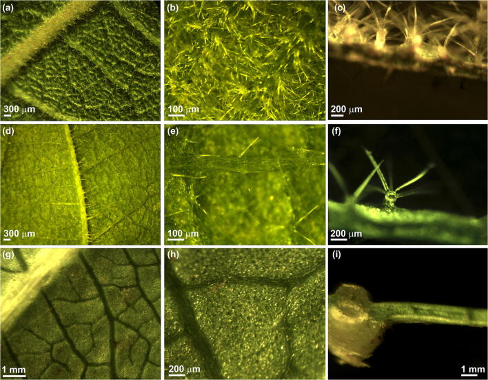
Fig. 2.
Leaf structure in different Actinidia species: A. chinensis var. chinensis (a, b and c), A. chinensis var. deliciosa (d, e and f) and A. arguta (g, h and i). a Abaxial surface of A. chinensis var. chinensis leaf showing a lateral vein. b Higher magnification of A. chinensis var. chinensis leaf surface showing a dense layer of stellate trichomes. c Transversal section of a A. chinensis var. chinensis leaf. The abaxial surface is on top of the image, and it is characterized by petiolate stellate trichomes. On the adaxial surface, uniseriate multicellular trichomes are visible. d Abaxial surface of A. chinensis var. deliciosa leaf showing a lateral vein. e Trichome distribution in A. chinensis var. deliciosa abaxial leaf surface showing stellate trichomes. f Transversal section of an A. chinensis var. deliciosa leaf. g Abaxial surface of an A. arguta leaf showing a lateral vein. h Higher magnification of the abaxial surface of an A. arguta leaf characterized by the absence of trichomes. i Transversal section of A. arguta leaf. The abaxial surface is on the top

