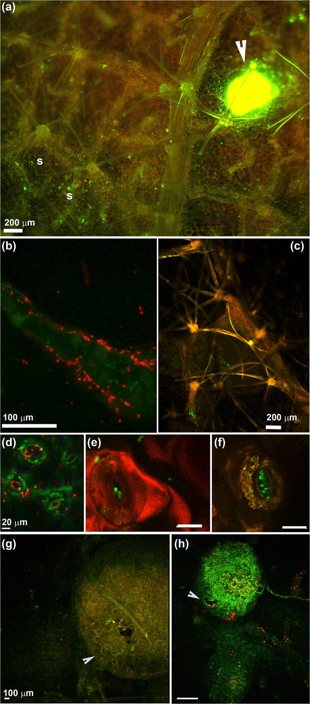
Fig. 3.
Kiwifruit leaves (a–f) and lenticels (g–h) colonized by Psa CFBP7286-GFPuv (a, c, e, f) or CFBP7286-DsRED (b, d, h). a Fluorescence stereomicroscope photograph of the abaxial surface of an A. chinensis var. deliciosa leaf. Several infected stomata are highlighted as bright green fluorescent spots. The arrow indicates a leaf lesion producing a drop of bacterial exudate. Stomata are highlighted with an ‘s’. b Uniseriate multicellular trichome colonized by Psa. Each red rod is a bacterial cell. Psa forms microcolonies at the base of the trichome. c Abaxial surface of a A. chinensis var. chinensis leaf. The base of several petiolate stellate trichomes is colonized by Psa CFBP7286-GFPuv (bright green signal). d–e Confocal laser scanning (CLSM) micrographs of A. chinensis var. deliciosa and A. chinensis var. chinensis (f) showing colonization of stomata by Pseudomonas syringae pv. actinidiae. To visualize the host colonization in vivo, the strains CFBP7286-DsRED (d) or CFBP7286-GFPuv (e, f), expressing a red or green fluorescent protein, were used. In micrograph (d) each red rod is a single pathogen cell; in (e) and (f), Psa cells are visualized as green rods. Unless specified, measuring bars in the same raw have the same length. g Confocal laser scanning micrograph of a healthy lenticel of a 2-week-old A. chinensis var. chinensis shoot. The lenticel surface is characterized by stomata-like structure indicated by the arrow. In the central part of the lenticel, a dark micro lesion, due to the swelling of lenticel in high humidity conditions, is also visible. h Lenticel infected by Psa CFBP7286-DsRED. Each red spot is a bacterial microcolony. Psa colonize the lenticel surface and it is associate to a microlesion due to lenticel swelling (arrow)

