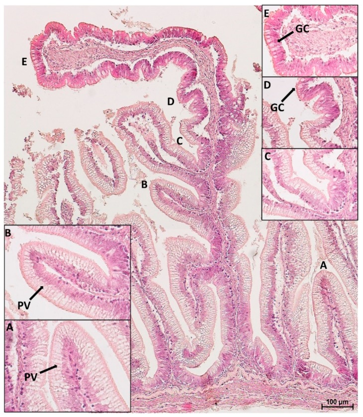
Figure 4.
HE stained section of a complex fold in the distal intestine: parietal villi (A), as well as villi emerging from the basal part of the fold (B,C), were both covered by pinocytotic vacuoles (PV). At one point, the epithelium morphology along the fold, drastically changed: pinocytotic vacuoles tended to disappear (D). The apex of the fold presented goblet cells (GC) and non-vacuolated enterocytes (E).

