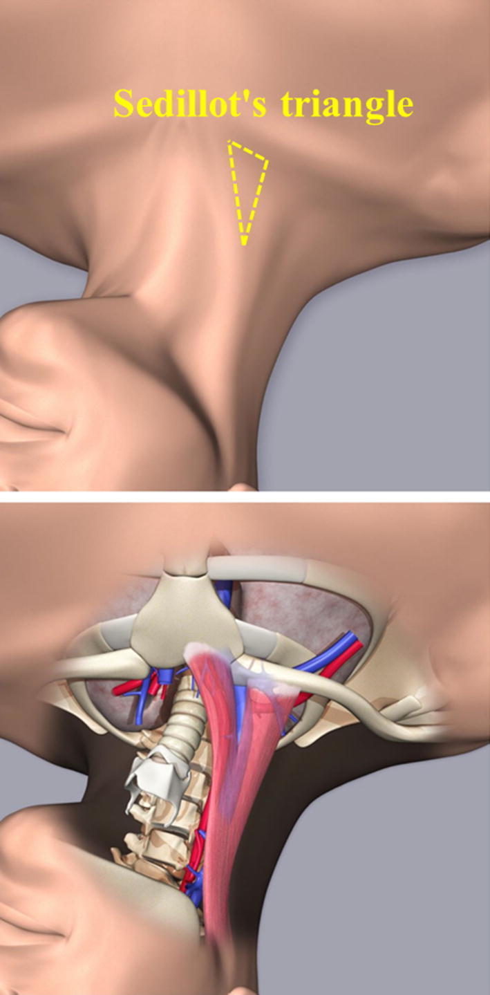
Fig. 1.

Sedillot’s triangle. Sedillot’s triangle comprises the sternocleidomastoid muscle and the clavicle. The triangle can be visually approximated or palpated. In most cases, the internal jugular vein is located within the triangle. Furthermore, insertion and catheterization within the triangle have the benefit of easy handling of the needle and dilator because of the thin tissue layer between the skin surface and the vein
