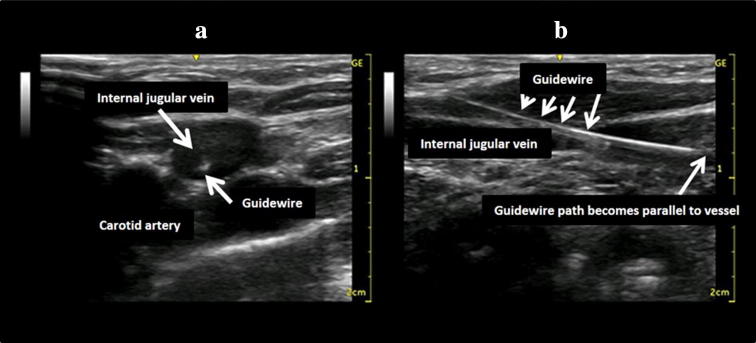
Fig. 15.
Confirmation of guidewire location using ultrasound images. The patient was a 6-month-old infant. The guidewire can be seen inside the internal jugular vein in the short-axis image (a). However, this alone cannot determine if the guidewire tip is inside the vein. The long-axis image shows that the guidewire path gradually becomes parallel to the vessel wall (b)

