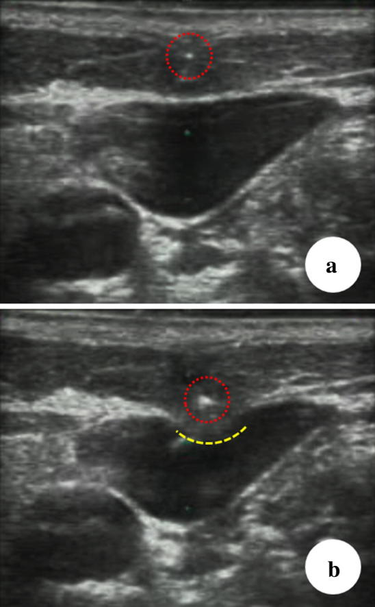
Fig. 5.

Pitfall of the short-axis out-of-plane technique. The yellow dotted line shows the dimple-like deformed anterior vein wall. It indicates the needle reaching and pushing the anterior vein wall. The white dot represents the shaft of the needle. a The needle tip (dotted red circle) is on the ultrasound beam. b The ultrasound image depicts part of the needle shaft (dotted red circle)
