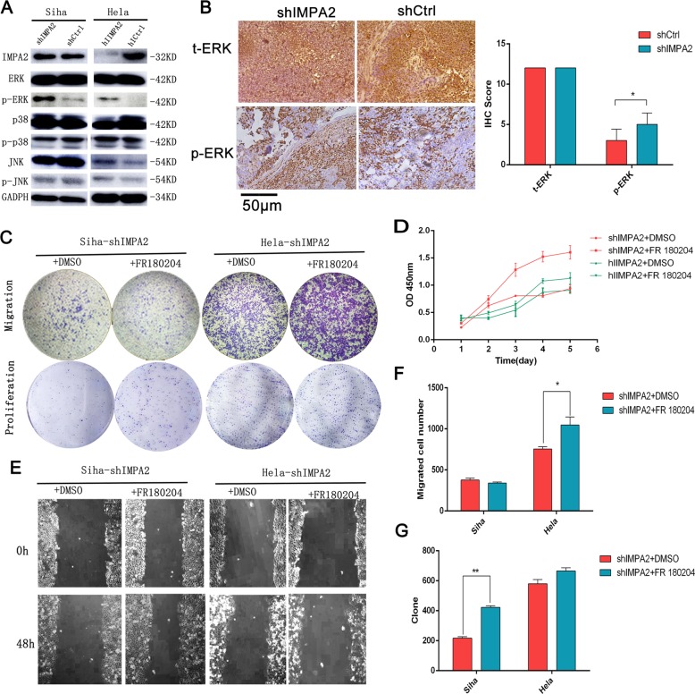
Fig. 6. Silenced IMPA2 activated ERK and the IMPA2-induced cell proliferation and migration were revised by ERK inhibitor.
a The expression levels of IMPA2 and MAPK signaling molecules ERK, p-ERK, p38, p-p38, JNK, p-JNK, and GAPDH protein in IMPA2 silencing SiHa or HeLa cells were detected by western blotting. b ERK and p-ERK expression of the inxenografts were detected by immunohistochemical staining and the immunoreactivity scores of t-ERK and p-ERK were shown. Scale bar, 50 μm. The migratory and proliferation potential of IMPA2 silencing SiHa or HeLa cells treated with FR 180204 or DMSO were analyzed by the transwell cell migration and colony formation assay (c). CCK-8 assay (d), wound healing assay (e). Number of migratory cells (f) and clone (g) was shown as mean ± SD from three independent experiments using triplicate measurements and statistically analyzed with Student’s t-test in each experiment. *p < 0.05, **p < 0.01.

