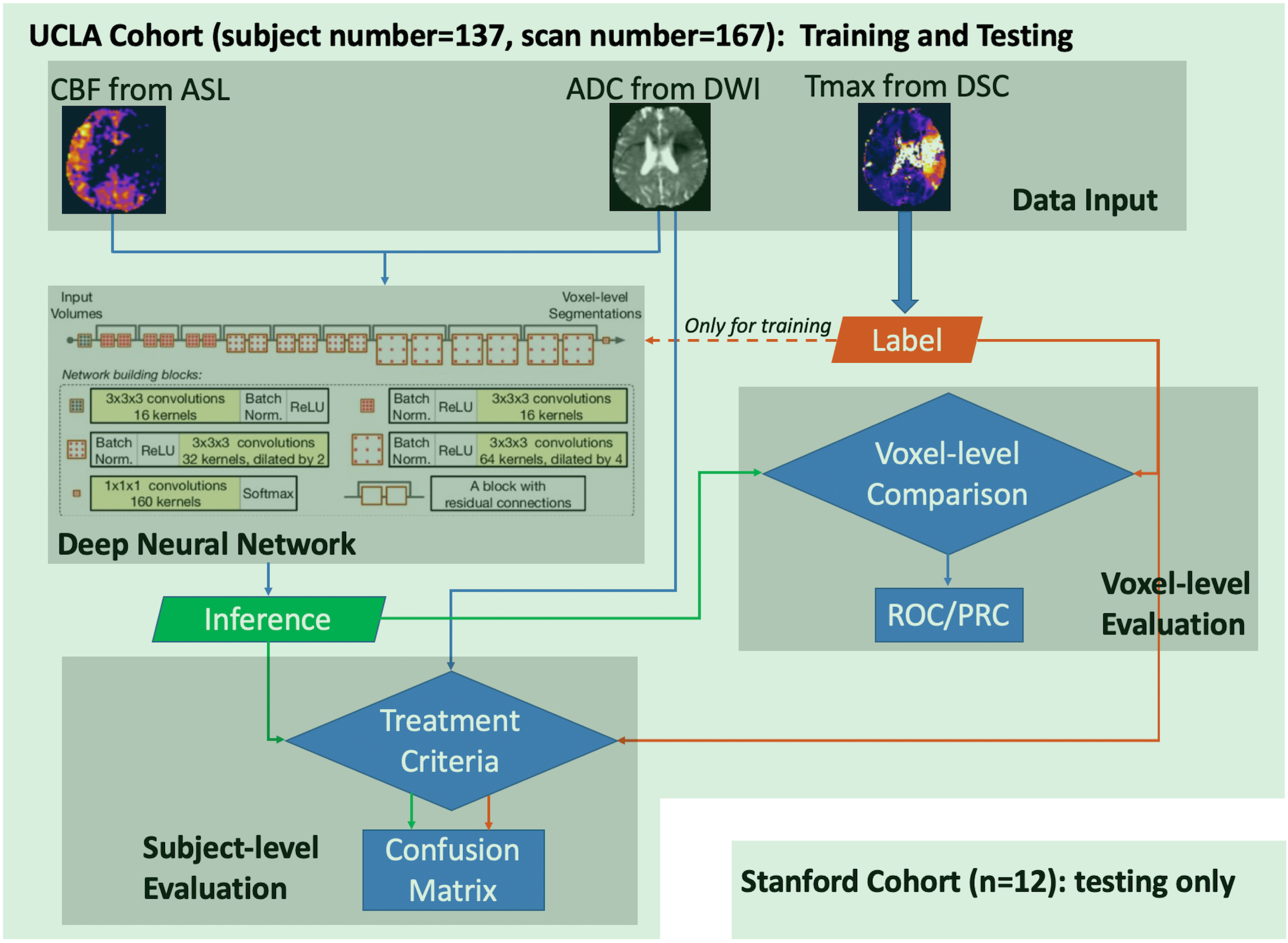
Figure 1. Flowchart of the training and evaluation of DL model.

DSC MRI scans were first processed to generate parameter maps (CBF, ADC, and Tmax), then the data were used to train the Deep Neural Network. After training, inference was made and evaluated at voxel-level and subject-level. The UCLA cohort was used for both training and testing, while the Stanford cohort was only used for testing purposes.
