Figure 3.
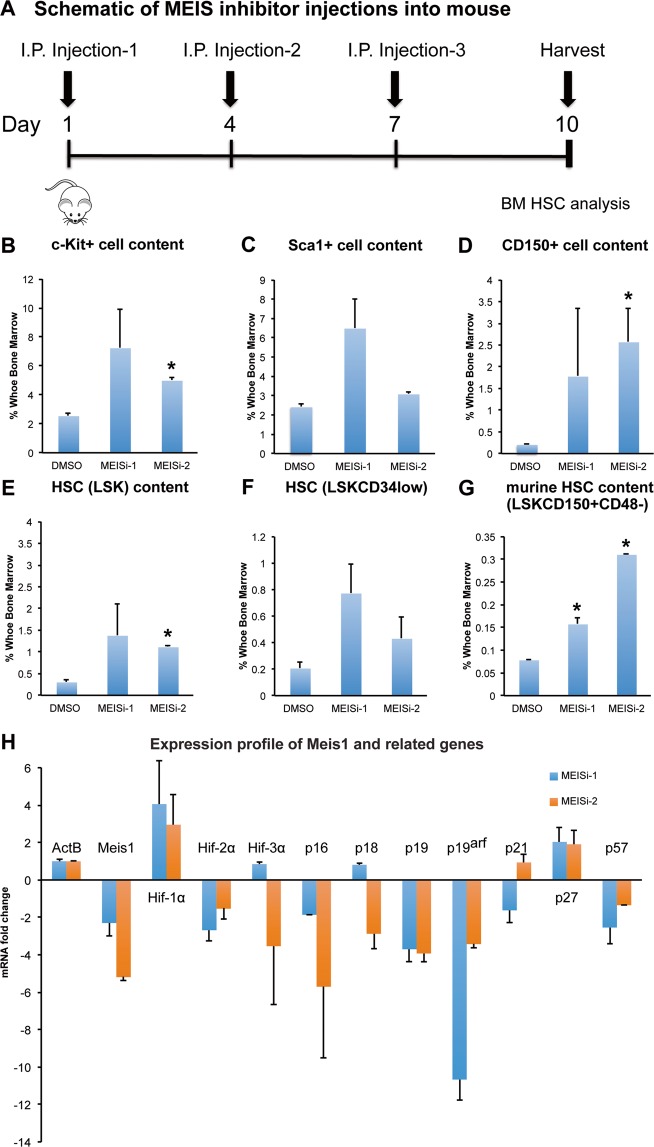
Phenotypical HSC antigen analysis post MEISi injections. (A) Schematic showing the intraperitoneal injections of MEIS inhibitors and BM analysis at day 10. In vivo analysis of HSC compartment post MEISi treatments was carried out by analysis of (B) c-Kit+, (C) Sca-1+ cell content, (D) CD150+ cell content, (E) LSK cell content, (F) LSKCD34low cell content and (G) LSKCD48−CD150+ HSC content in the whole bone marrow following injection of MEISi-1, MEISi-2 and DMSO control. (H) Expression profile of Meis1 and related target genes post MEISi injections in the whole bone marrow cells. n = 3, *p < 0.05.

