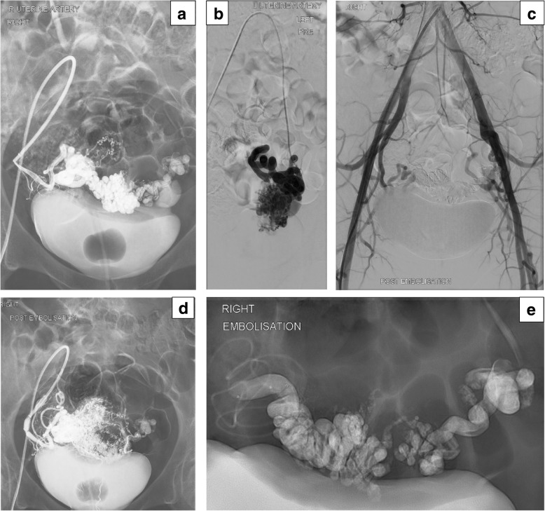
Fig. 2.
a-e. 41-year-old female with symptomatic acquired uterine AVM. a Right uterine artery demonstrates hypertrophied right uterine artery and AV shunting, with draining venous varix; b Left uterine artery with evidence of AV shunting and a well demonstrated nidus; c Aortogram post bilateral UAE, showing obliteration of the AV shunting and no further feeder from internal and / or external iliac arteries; d Post right uterine artery embolisation with PHIL, showing no evidence of AV shunting. Adequate nidus penetration with the embolic cast also seen; e Post bilateral UAE demonstrating PHIL cast and the Scepter XC catheter. It also nicely demonstrates the marked tortuosity of the hypertrophied right UA

