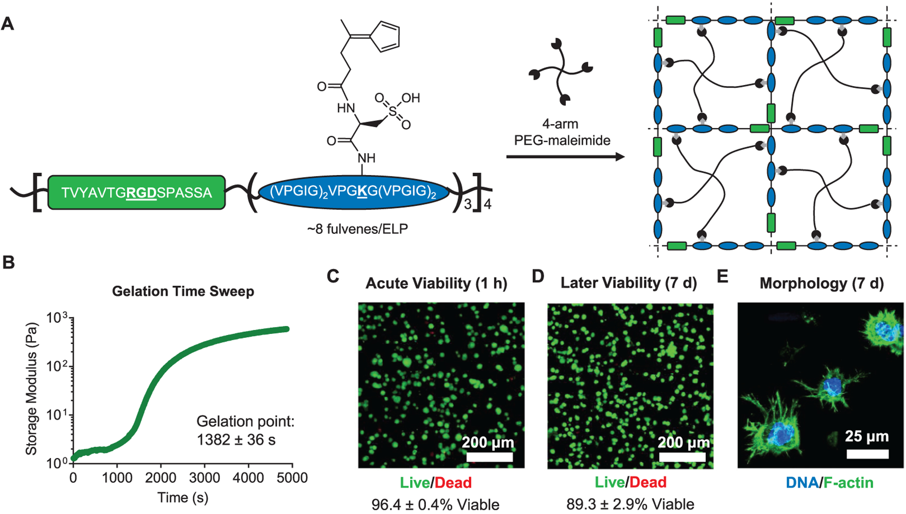
Figure 5.

Engineered protein–PEG hybrid DA hydrogels support the culture of hMSCs. (A) Schematic of fulvene-modified ELPs containing a cell-adhesive RGD domain (green) and a structural elastin-like domain (blue). Upon mixing with a 4-arm PEG–maleimide crosslinker, the fulvene ELPs form hydrogel networks. (B) Gelation time sweep for ELP–PEG fulvene DA gels. Gelation point data are mean ± s.d., n = 3. Viability of hMSCs encapsulated in ELP–PEG fulvene DA gels after (C) 1 h and (D) 7 days, measured by a live/dead cytotoxicity assay. Viability data are mean ± s.d., n = 4. (E) Confocal fluorescence micrograph showing spreading of hMSCs cultured in ELP–PEG fulvene DA gels for 7 days. The actin cytoskeleton was stained with phalloidin (green) and the nuclei were stained with Hoechst (blue).
