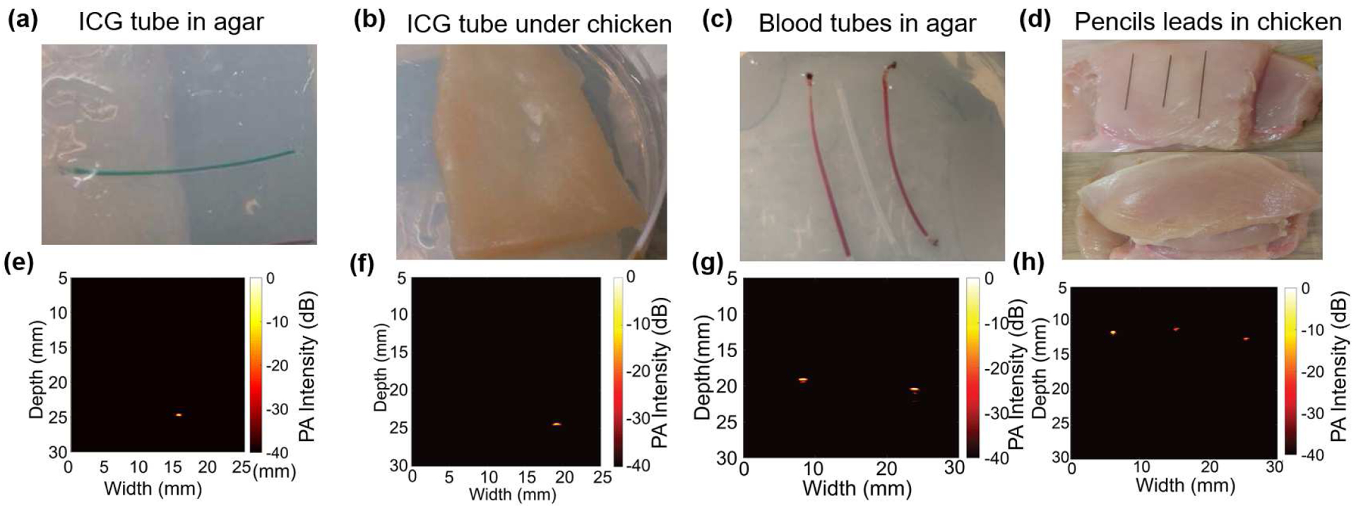
Fig. 9.

B-mode photoacoustic imaging (PAI): (a) A clear agar-gel phantom with an ICG-filled tube as target. (b) Phantom shown in (a) is covered with 2 mm thick chicken tissue layer to introduce scattering of the light. (c) A clear agar-gel phantom with two blood filled polymer tubes embedded at 5 mm depth with an unfilled tube placed between the two tubes. (d) Pictures showing the preparation of chicken tissue-based phantom. Three 0.3 mm pencil leads were placed on the chicken bed and then an 8 mm thick chicken tissue layer was placed on top of the pencil leads. (e), (f), (g), and (h) are the reconstructed B-mode (photoacoustic images from phantoms shown in (a), (b), (c), and (d) respectively. The color-bar for all the reconstructed images represents the photoacoustic intensity, normalized with respect to the maxima within each image, on log scale with 0 dB to Š40 dB range. All images were acquired using linear scanning of single element PMUT with 0.1 mm step size.
