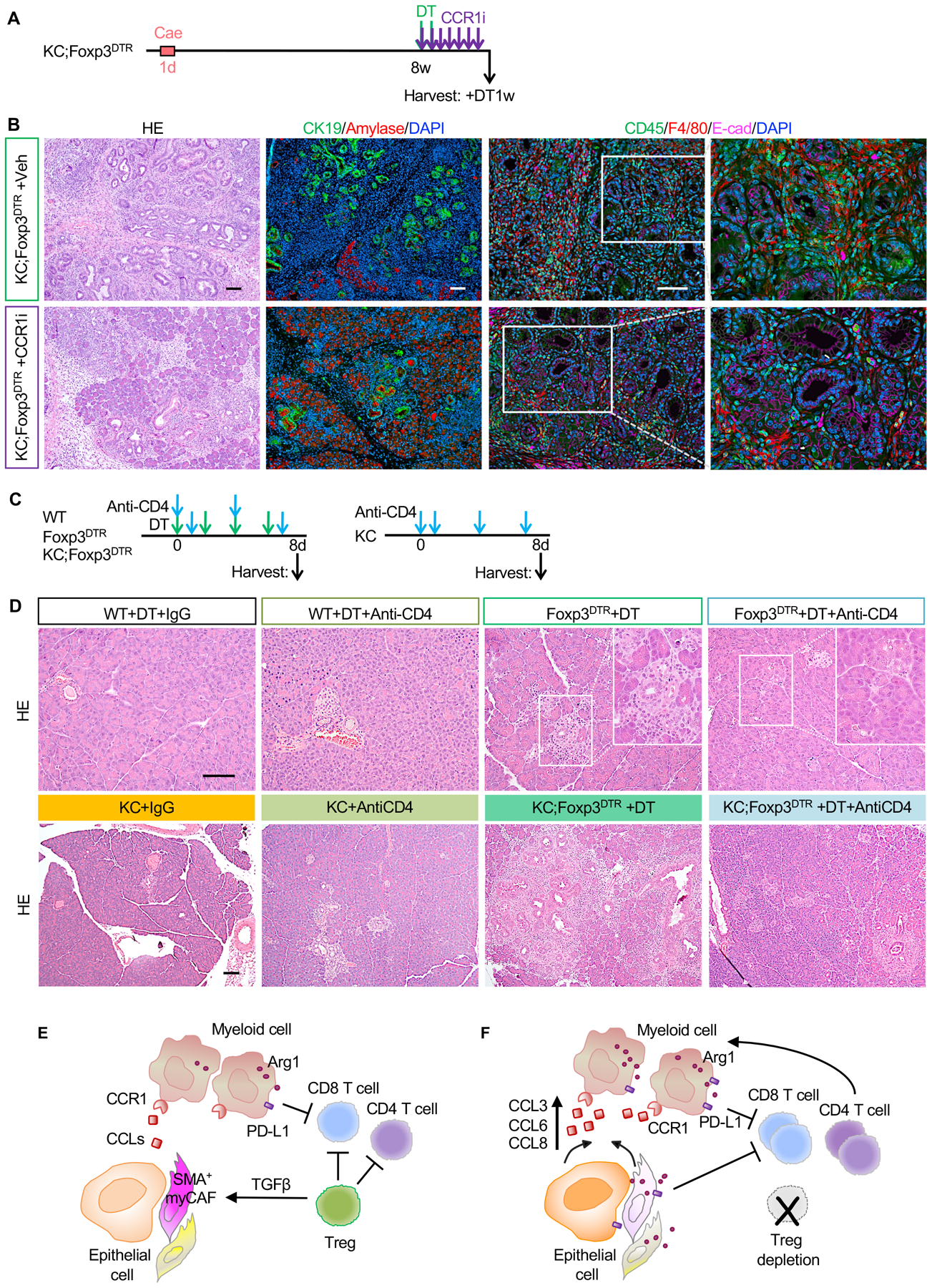
Figure 7. CCR1 inhibition and CD4+ T cell depletion abrogate Treg depletion induced PanIN progression.

(A) Experimental design, n=5–6 mice/cohort. (B) H&E staining, co-immunofluorescent staining for CK19 (green), Amylase (red) and DAPI (blue), and co-immunofluorescent staining for CD45 (green), F4/80 (red), E-cad (magenta) and DAPI (blue) in control and CCR1 inhibitor treated KC;Foxp3DTR pancreata. Scale bar 100 μm. (C) Experimental design and (D) H&E staining for WT, Foxp3DTR, KC and KC;Foxp3DTR mice that received DT treatment and/or anti-CD4 antibody. n=3–4 mice/cohort. Scale bar 100 μm. (E) Working model. Tregs inhibit CD8+ T cells, but also restrain pathogenic CD4+ T cell responses. Tregs regulate the production of TGFβ thus promoting the differentiation of Smooth Muscle Actin (SMA)+ fibroblasts (myCAFs). (F) Treg depletion results in loss of TGFβ and compensatory changes in the fibroblast population that in turn secrete myeloid-recruiting chemokines, driving increased recruitment of myeloid cells and failing to alleviate immunosuppression. Treg depletion also unleashes pathogenic CD4+ T cell responses that promote pancreatic inflammation and carcinogenesis.
