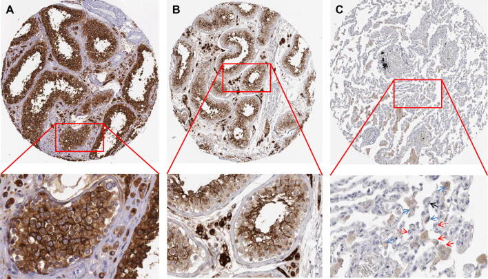
Fig. 3.
Immunohistochemistry (IHC) images of normal tissues (testis and lung) for ACE2. a IHC image of normal testis tissue from a male of age 38 (Patient id: 305). b IHC image of normal testis tissue from a male of age 26 (Patient id: 2254). c IHC image of normal lung tissue from a female of age 49 (Patient id: 2268). Bottom panels, enlarged pictures from top panels. Blue arrows indicate the representative positive results in macrophages, black arrow indicates the representative positive results in type I alveolar epithelial cells, red arrows indicate the representative positive results in type II alveolar epithelial cells (c, red arrow), but dashed red arrows indicate the representative negative staining of type II alveolar epithelial cells. (Color figure online)

