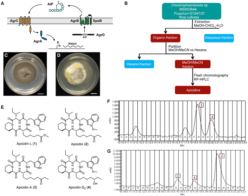
Figure 1. The agr System and Isolation of Apicidin.
(A) Schematic of the agr system.
(B) Flowchart for isolation of apicidins from solid-state culture extracts.
(C) Chaetosphaeriaceae sp. (MSX53644) grown on PDA (scale bar, 10 mm).
(D) Fusarium sp. (G134 and G137) grown on PDA (scale bar, 10 mm).
(E) Apicidin structures.
(F) Preparative chromatogram (λ= 254 nm) of the fraction used to purify compounds 1 and 2 from MSX53644.
(G) Preparative chromatogram (λ = 254 nm) of the fraction used to purify compounds 2–4 from G134 and G137 (each chromatogram is representative of multiple runs).

