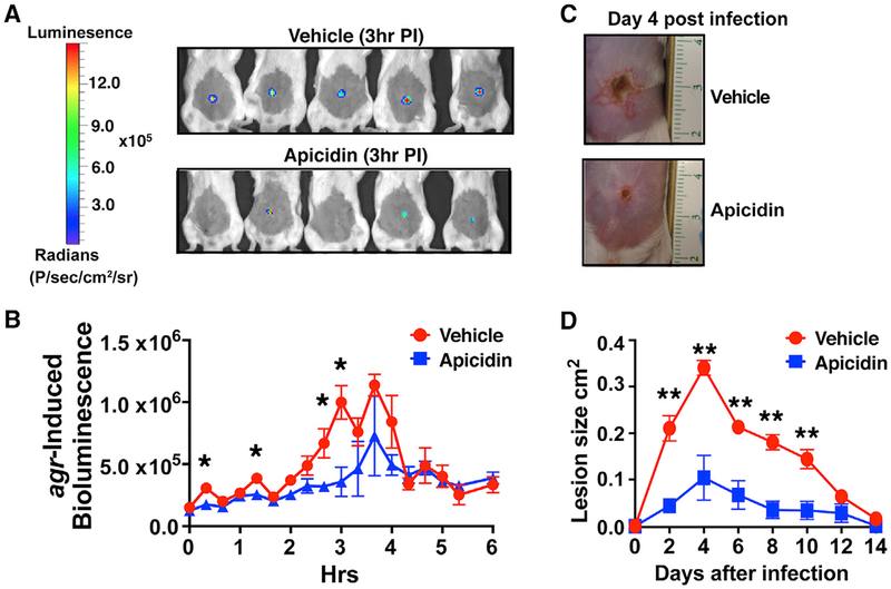
Figure 4. Apicidin-Mediated Quorum-Sensing Inhibition Corresponds with Atten uated Skin Injury.
(A) Images of agr-P3 reporter activity (bioluminescence) 3 h post infection (±5 μg apicidin).
(B) Kinetics of agr activation in apicidin and vehicle-control-treated mice after infection (n = 5).
(C) Representative images of apicidin or control groups at the indicated time points after infection (n = 5).
(D) Skin lesion size measurements at the indicated time points after infection. Error bars represent SEM. Post-test *p < 0.05 and **p < 0.01.

