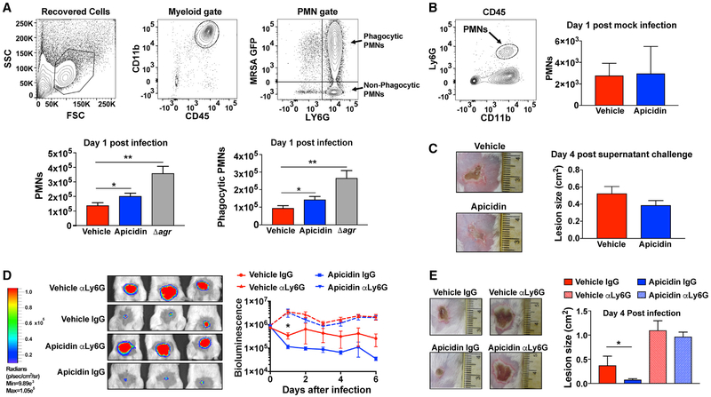
Figure 6. Apicidin Treatment Enhances PMN Responses after MRSA Skin Challenge.
(A) Gating strategies (top) for identification of PMNs recovered from lesional skin and corresponding PMN accumulation values (bottom) 1 day after intradermal challenge with 2 × 10⁷ MRSA WT expressing GFP (± apicidin) or its agr null counterpart (n = 8). Error bars represent SEM. Post-test *p < 0.05 and **p < 0.01.
(B) Gating strategy (left) and enumerated PMNs (right) 1 day after intradermal saline challenge (± apicidin; n = 8).
(C) Representative skin injuries 4 days after sterile challenge (intradermal) with 30 μL of WT MRSA supernatant at the skin sites intradermally inoculated with saline (± apicidin) 3 h prior (n = 6).
(D) Representative bioluminescence images 1 day after intradermal challenge (2 × 10⁷ CFUs MRSA Lux+) among apicidin-treated and control groups receiving 100 μg of the PMN-depleting anti-Ly6G antibody or isotype control via intraperitoneal injection administered both the day prior and the day of infection. Corresponding time course values (right) of MRSA burden for the indicated groups (n = 6).
(E) Representative images of skin injury (left) and corresponding lesion size measurements 4 days after infection for the indicated groups (n = 5). Error bars represent SEM. Post-test *p < 0.05 and **p < 0.01.

