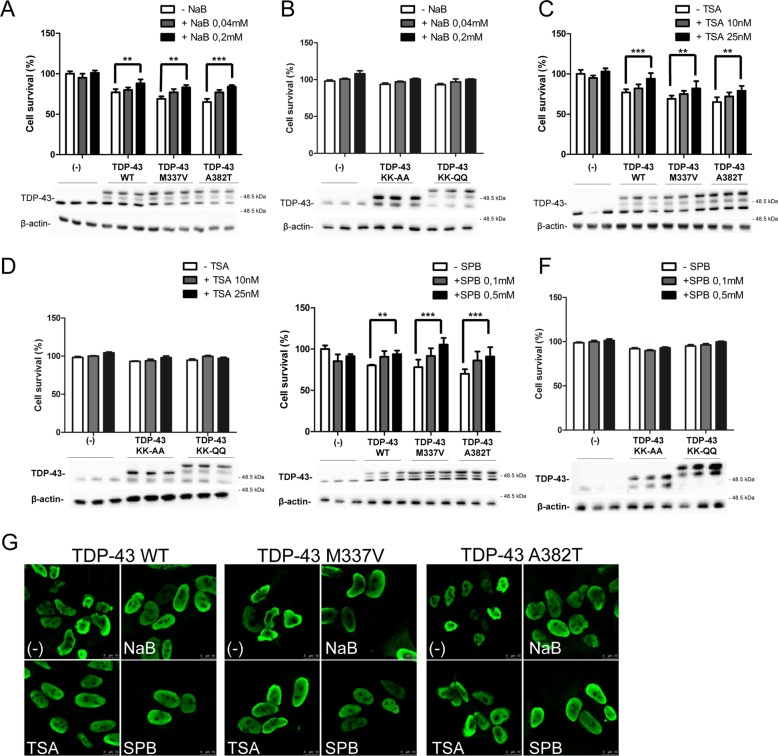
Fig. 4. Effect of HDACi on TDP-43-induced cell toxicity in SH-SY5Y cells.
MTS assay on SH-SY5Y cells transduced with the indicated adenoviral particles encoding for TDP-43 WT, M337V, A382T, KK-AA, or KK-QQ (MOI 10 pfu/cell), and treated whit different HDACi. Forty-eight hours after transduction and concomitant HDACi treatment cell viability was assessed by a colorimetric assay. At the end of the assay the cell extracts of the four replicates for each time point were pooled and analysed by western blot using anti-TDP-43 antibody. anti-βactin was used as loading control. NaB 0.04 and 0.2 mM (a, b); TSA 10 nM and 25 nM (c, d); SPB 0,1 mM and 0,5 mM (e, f). The data were obtained from four independent experiments; **p > 0.01 and ***p > 0.001 analysed by using one-way ANOVA with Bonferroni’s Multiple comparison post-hoc test (g) Immunofluorescence on SH-SY5Y transduced with different myc-tagged-TDP-43 isoforms treated for 48 h with NaB 0.2 mM, TSA 25 nM, and SPB 0.5 mM. Cells were labeled with an anti-Myc antibody and detected with a secondary conjugate to Alexa 488 anti-mouse fluorophore as a secondary antibody. The slides were analysed by Leica confocal microscope. Scale bars = 10 μm.

