
Fig. 2.
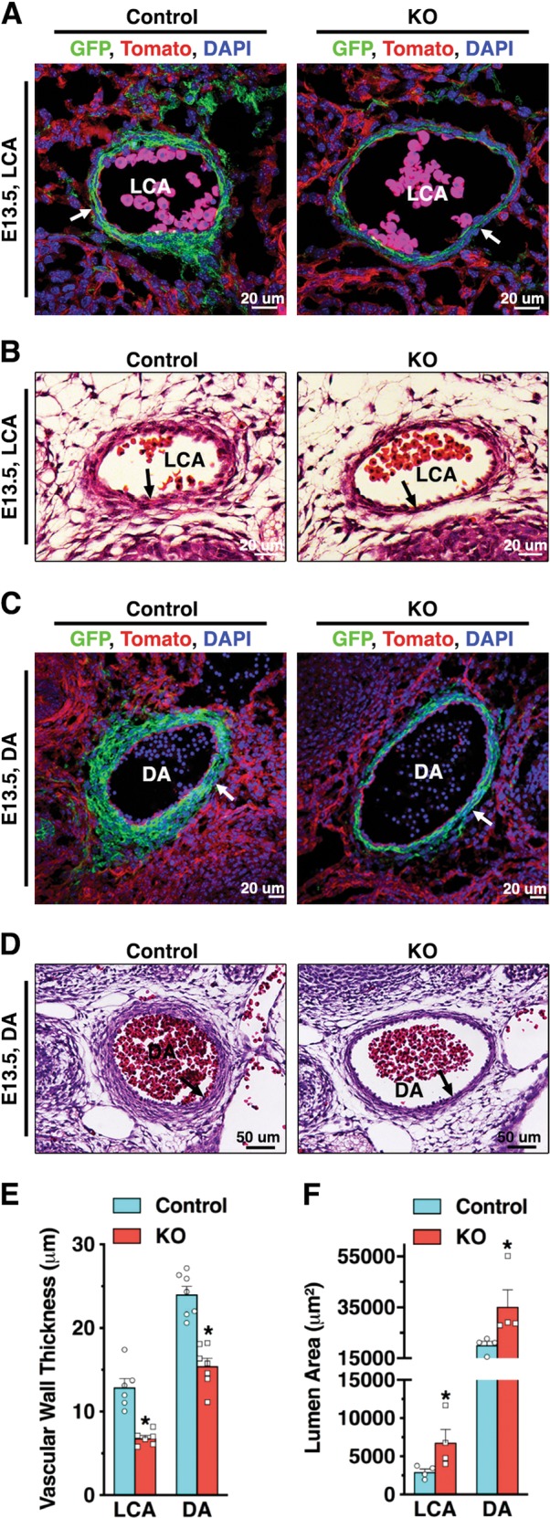
Conditional deletion of Tead1 in VSMCs leads to an atrophic arterial wall and dilated arterial lumen. At E13.5, Tagln-Cre+/Tead1F/W/mTmG+/− heterozygous control and Tagln-Cre+/TEAD1F/F/mTmG+/− mutant embryos were harvested and sectioned for the direct visualization of GFP-marked VSMCs (a, c) or for HE staining (b, d) of the left common carotid artery (LCA, a, b) and dorsal aorta (DA, c, d). Thickness of the media wall (e) and total lumen area (f) were quantified from the cross sections shown in panels (a) and (c). *p < 0.05