Fig. 3.
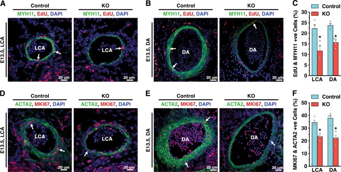
Tead1 KO embryos display proliferative defects in VSMCs. Pregnant dams carrying E13.5 control or Tead1 KO embryos were peritoneally injected with EdU to mark proliferating cells for 2 h. Embryos were then isolated, embedded and sectioned for immunostaining with antibodies for the smooth muscle differentiation marker MYH11 (green) together staining with the incorporated EdU (red) in LCA (a) or DA (b). Samples were counter-stained with DAPI (blue) to visualize nuclei. The percentage of EdU-positive VSMCs in the arterial wall of LCA or DA were measured and graphed as shown in panel (c). Similarly, sections were stained with antibodies for the smooth muscle differentiation marker ACTA2 (green) and the marker of cell proliferation, MKI67 (red) in LCA (d) or DA (e), respectively. The relative number of MKI67-positive VSMCs was measured and is shown in (f). N = 4 embryos per genotype. *p < 0.05

